
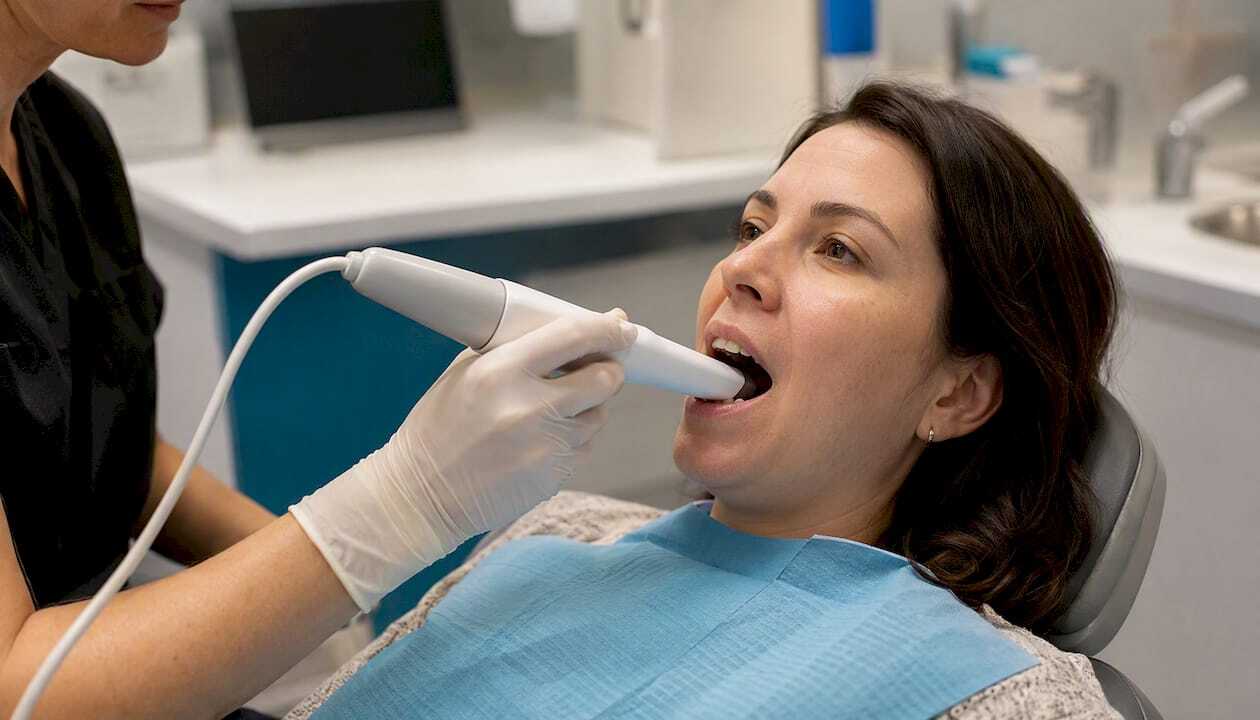
Digital dental impressions use a small handheld wand called an intraoral scanner to capture thousands of images per second, which the software merges into a precise 3D virtual model of your teeth and gums. No putty trays, no gagging, no waiting for material to set. For most single-tooth restorations, short-span bridges, and orthodontic records, digital impressions are clinically accurate and more comfortable than traditional methods. Some full-arch implant cases and deep-margin situations still favor conventional impressions, but those are the exceptions rather than the rule.
Key Takeaways
Digital dental impressions are clinically accurate for single-tooth and short-span restorations, more comfortable than putty trays for most patients, and increasingly available in U.S. dental practices, though full-arch accuracy and operator skill remain important variables.
| Point | Details |
|---|---|
| What they are | An intraoral scanner captures a real-time 3D model of your teeth, replacing putty trays entirely. |
| Strongest evidence base | Single-tooth restorations and short-span bridges show consistently high accuracy across major scanner families. |
| Main limitations | Deep subgingival margins, movable soft tissues, and some full-arch implant cases may still require conventional impressions. |
| Two questions to ask | Ask which scanner your dentist uses and whether the workflow allows same-day or faster restoration delivery. |
| Local option | Integrativedentalofdenver uses digital scanning within an airway-centered care model for orthodontic, restorative, and appliance cases. |
Table of Contents
- How digital dental impressions work at your appointment
- What clinical evidence says about intraoral scanner accuracy
- Key patient benefits and common clinical uses
- When your dentist may still choose a conventional impression
- How much digital impressions cost and where to find them
- Why operator skill shapes your scan outcome
- Questions to ask your dentist about digital impressions
- Insurance coverage and reimbursement for digital impressions
- Timeline from impression to restoration or appliance delivery
- A clinical perspective on digital impressions and airway-centered care
- Integrativedentalofdenver offers digital scanning as part of whole-health dental care
- Sources
How digital dental impressions work at your appointment
The entire process is more straightforward than most patients expect. The scanner wand, roughly the size of an electric toothbrush, captures real-time 3D data that appears on a chairside monitor as the clinician moves it across your teeth. You can watch your own dental anatomy build on screen, which many patients find reassuring.
Here is the typical appointment flow:
- Preparation. The clinician dries the teeth lightly with air and may apply a thin anti-reflective powder to highly polished surfaces (many modern scanners skip this step entirely).
- Occlusal scan. The wand passes over the biting surfaces first, establishing the arch shape.
- Lingual scan. The scanner captures the tongue-side surfaces.
- Buccal scan. The cheek-side surfaces are recorded, completing the arch.
- Bite registration. You close naturally while the scanner records how upper and lower teeth meet.
- Review. The clinician inspects the 3D model on screen, fills any gaps with a quick rescan, and confirms margin quality.
- Transmission. The file goes directly to the dental lab or in-office CAD/CAM milling unit, often within minutes.
The scanner families you are most likely to encounter include the 3Shape Trios series, iTero (by Align Technology), Medit (such as the Medit i500), Planmeca (including the Emerald and other Planmeca scanners), and Dentsply Sirona (the Primescan and CEREC family). Each uses slightly different optical technology, but the patient experience is similar across all of them.
Hygiene between patients follows strict protocols: scanner tips are either single-use disposable sleeves or autoclavable components, and the wand body is wiped with hospital-grade disinfectant.
Pro Tip: Keep your lips and tongue relaxed and breathe through your nose during the scan. If you feel uncomfortable at any point, raise your hand. The clinician can pause, let you rest, and resume without losing the data already captured.
What clinical evidence says about intraoral scanner accuracy
Digital impressions are clinically acceptable and often equal or superior to conventional impressions for single-tooth and short-span restorations. Full-arch accuracy is more variable and depends heavily on scanner model, scan pattern, and operator experience.
A narrative review published in PMC/NIH confirmed that intraoral scanners achieve high accuracy for single-tooth restorations and improve both workflow efficiency and patient comfort, while noting that movable soft tissues, deep subgingival margins, and long-span prostheses remain areas of limitation. A comparative in vitro study of ten intraoral scanners found that trueness (how close a scan is to the true shape) varied among scanner models, while precision (repeatability) was generally similar across devices. That same study noted that trueness decreases as scan distance increases, which is why careful, consistent scan patterns matter for full-arch work.
For full-arch cases specifically, a systematic review and meta-analysis concluded that intraoral scanning accuracy often falls within clinically acceptable thresholds but flagged significant heterogeneity among studies, making sweeping conclusions difficult. The practical takeaway: full-arch digital impressions can work well, but the outcome depends more on technique and scanner selection than single-tooth scans do.
| Dimension | Single tooth / short span | Full arch |
|---|---|---|
| Trueness and precision | Consistently high across major scanner families | Variable; scan pattern and operator technique are critical |
| Best clinical indications | Crowns, inlays, onlays, short bridges, implant crowns | Orthodontic records, complete dentures (selected cases), implant planning with scan bodies |
| Scan speed and chair time | Typically faster than conventional impressions | Longer; more passes required |
| CAD/CAM integration | Direct to in-office mill or lab | Direct to lab; some full-arch cases still use hybrid workflows |
| Powder or special prep | Rarely needed with current scanners | Occasionally needed for highly reflective surfaces |
| Operator dependency | Moderate | High; consistent scan path reduces stitching errors |
The 3Shape Trios, iTero, Medit, Planmeca, and Dentsply Sirona Primescan all appear in published scanner-comparison literature. No single device dominates every accuracy metric across all clinical scenarios, which is why asking your clinician about their specific scanner and case volume matters more than brand loyalty.
Key patient benefits and common clinical uses
The shift to digital scanning offers patients several concrete advantages over traditional putty impressions.
Patient-facing benefits:
- No putty tray pressed into your mouth, which significantly reduces gag reflex triggers
- Faster appointment time in most single-tooth and short-span cases, as confirmed by patient perception research showing higher comfort and preference for digital methods
- Immediate on-screen visualization of your teeth, allowing you to see what the dentist sees
- Digital records stored securely and retrievable for future treatment planning without a new impression
- Fewer retakes compared to conventional impressions, where a single bubble or distortion requires starting over
- Direct integration with dental CAD/CAM systems for same-day chairside restorations in equipped practices
- Reduced material waste and no need to disinfect or ship physical plaster models to a lab
Common clinical applications:
- Single crowns, inlays, and onlays (the strongest evidence base)
- Short-span fixed bridges (two to three units)
- Implant crowns using scan bodies placed on the implant fixture
- Orthodontic records and digital model fabrication for clear aligner therapy (such as Invisalign)
- Occlusal splints and night guards
- Palate expanders and other removable appliances
- Chairside same-day restorations when the practice has an in-office milling unit
For patients at Integrativedentalofdenver who are pursuing airway-focused orthodontics or appliance-based treatments, digital scans also support precise fabrication of palatal expanders and oral appliances without the discomfort of conventional trays.
When your dentist may still choose a conventional impression
Digital impressions are not the right tool for every clinical situation. Understanding the exceptions prevents surprises at your appointment.
Main clinical limitations:
- Deep subgingival margins. When a crown preparation extends well below the gumline, the scanner cannot reliably capture the margin without soft-tissue management (retraction cord or laser). In some cases, a conventional impression after retraction still produces a cleaner result.
- Movable soft tissues. Fully edentulous arches with mobile vestibular tissue are difficult to scan accurately because the tissue shifts between frames, creating stitching errors in the 3D model.
- Very long-span prostheses. Some full-arch implant-supported bridges require dimensional accuracy that current intraoral scanners cannot consistently achieve across the full span; conventional impressions or scan-body workflows with desktop scanners remain common here.
- Post-and-core preparations. Deep canal preparations for post placements are often better captured with conventional methods.
Operator- and equipment-related factors that increase rescans:
- Inconsistent scan path (skipping the occlusal-to-lingual-to-buccal sequence)
- Excessive saliva or blood in the field obscuring the scanner’s optical view
- Undercuts in the preparation that the scanner’s angle cannot reach
- Outdated scanner software or an uncalibrated device
Hybrid workflows address many of these gaps. A clinician might take a conventional impression for a challenging case and then digitize it on a desktop scanner, or use scan bodies placed on implant fixtures to capture implant positions digitally even when the surrounding tissue is difficult to scan directly.
How much digital impressions cost and where to find them
Many dental practices across the United States now offer intraoral scanners, though adoption is not universal. The practice investment for a scanner system is typically substantial, varying considerably depending on brand and software package, which is why smaller or rural practices may not yet have one.
From a patient’s perspective, the cost of a digital impression is usually bundled into the procedure fee rather than billed as a separate line item. A crown, for example, carries the same CDT billing code whether the impression was taken digitally or with putty. You are unlikely to see a separate charge labeled “digital impression fee” on your explanation of benefits.
What to check before your appointment:
- Ask whether the practice has an in-office milling unit (CAD/CAM) or sends files to an external lab, since this affects turnaround time.
- Confirm with your insurer whether the procedure code your dentist will use is covered under your plan; the impression method itself rarely changes coverage.
- Ask whether the digital workflow changes the number of appointments needed (some same-day crown workflows eliminate a second visit entirely).
Pro Tip: Ask your dentist specifically whether using a digital scan will shorten your restoration timeline. In practices with in-office CAD/CAM milling, a crown can sometimes be designed, milled, and seated in a single appointment, eliminating the temporary crown and the second visit.
Why operator skill shapes your scan outcome
Clinician experience and scanning technique are among the most significant variables in intraoral scanning accuracy. The scanner is a tool; the outcome depends on the person using it.
A 2023 PubMed review specifically identified the “operator effect” as a critical variable, noting that clinician experience, scan strategy, and device calibration significantly influence scan accuracy. Technology alone does not guarantee a perfect result.
What the research shows about operator dependency:
- Consistent scan patterns (occlusal surface first, then lingual, then buccal) reduce stitching errors, particularly in full-arch scans.
- Clinicians who perform higher scan volumes tend to produce more accurate full-arch results, reflecting a learning curve that flattens with practice.
- Scanner calibration and software version affect trueness; a well-maintained, regularly updated system outperforms a neglected one regardless of brand.
- Staff training programs offered by 3Shape, Align Technology (iTero), Medit, Planmeca, and Dentsply Sirona all include scan-pattern protocols, but individual clinician adherence varies.
When a practice claims superior accuracy for a specific scanner, that claim should be supported by the scanner model, software version, and scan pattern used. A result achieved under ideal in vitro conditions does not automatically transfer to a busy clinical environment.
Pro Tip: When you ask your dental team about their digital impression experience, request specifics: how many scans they perform per month and whether staff have completed the scanner manufacturer’s training certification. A confident, specific answer is a good sign.
Questions to ask your dentist about digital impressions
Use these questions at your consultation or treatment planning appointment to clarify what to expect.
Checklist:
- Which scanner do you use, and has your team completed manufacturer training?
- Will you use a direct digital scan, or is a hybrid workflow (scanning a conventional impression) more appropriate for my case?
- How long will the scan take, and will I need to hold still for extended periods?
- What happens if a rescan is needed? Is there an additional fee?
- Who reviews the final fit, and how are adjustments handled?
- Will my insurance cover this procedure, and does the impression method affect my coverage?
- What is the turnaround time from scan to delivery of my restoration or appliance?
Red flags to watch for:
- The practice cannot explain their lab workflow or who fabricates the restoration.
- Staff cannot describe their scanner training or how long they have been using the device.
- Multiple rescans occur without a clear explanation of why.
- No clear timeline is given for restoration delivery.
Insurance coverage and reimbursement for digital impressions
Insurance coverage for dental procedures in the United States is determined by CDT (Current Dental Terminology) procedure codes, not by the method used to take the impression. A crown preparation, for example, is billed under the same code whether the impression was digital or conventional. This means that in most cases, switching to a digital workflow does not change your out-of-pocket cost or your insurer’s reimbursement decision.
That said, a few nuances are worth understanding. Some practices charge a separate “digital records” or “digital scan” fee for orthodontic records or implant planning scans that fall outside a standard restorative procedure. These fees may or may not be covered depending on your plan. Always ask your dentist’s billing coordinator to confirm the specific codes being submitted before your appointment.
For patients using flexible spending accounts (FSAs) or health savings accounts (HSAs), dental procedure fees paid out of pocket are generally eligible expenses regardless of whether a digital or conventional impression was used. Verify with your plan administrator if you have questions about a specific procedure.
Timeline from impression to restoration or appliance delivery
One of the most practical advantages of digital impressions is the potential to compress the time between your scan and the delivery of your finished restoration or appliance.
Typical timelines by workflow type:
- Same-day chairside restoration (in-office CAD/CAM). In practices equipped with an in-office milling unit such as the CEREC system from Dentsply Sirona, a crown can be designed, milled from a ceramic block, and seated in a single appointment, often within two to three hours. No temporary crown is needed.
- Digital scan sent to an external lab. The file transmits electronically within minutes of the scan. Most dental labs return a finished crown or appliance within five to ten business days, comparable to or faster than the conventional impression-to-lab timeline, which also includes shipping time for physical models.
- Orthodontic aligners. Digital scans submitted to aligner manufacturers (for clear aligner therapy) typically result in a treatment plan preview within one to two weeks and aligner fabrication within two to four weeks after approval.
- Implant-supported restorations. Timelines vary based on healing stages, but the digital scan itself does not add time; it often reduces it by eliminating the need to pour and ship a stone model.
The key question to ask your dentist is whether their practice uses in-office milling or an external lab, since that single factor determines whether your restoration takes one appointment or two.
A clinical perspective on digital impressions and airway-centered care
At Integrativedentalofdenver, intraoral scanning is not simply a convenience upgrade. It is a functional part of how the clinic plans and delivers airway-centered orthodontic and restorative care. Digital 3D models allow Dr. Jim Bieneman and the clinical team to assess arch form, tooth position, and palatal dimensions with a level of detail that supports treatment planning for palate expansion, oral appliance therapy, and orthodontic correction simultaneously.
In a typical workflow at the clinic, a patient presenting for an orthodontic evaluation or a crown preparation receives a full-arch or quadrant scan that feeds directly into the treatment planning software. For orthodontic cases, the digital model integrates with the clinic’s broader assessment of airway dimensions and facial development, supporting the airway-first treatment principles that guide care across all age groups. For a restorative case such as a crown, the scan goes to the lab with the preparation design already confirmed on screen, reducing the chance of a remade restoration.
The approach reflects a broader philosophy: that precision at the impression stage prevents downstream problems in fit, function, and patient comfort. Digital workflows support that goal more reliably than conventional impressions do for the majority of cases the clinic handles.
Integrativedentalofdenver offers digital scanning as part of whole-health dental care
Patients in Denver who want the accuracy and comfort of intraoral scanning alongside a treatment plan that addresses breathing, sleep, and long-term oral health have a specific option worth considering. Integrativedentalofdenver uses digital scanning as part of its airway-centered approach to orthodontics, restorative care, and appliance therapy, meaning your scan data informs more than just a single crown or aligner tray.
Whether you are exploring adult airway orthodontics, a restorative procedure, or a first evaluation for a child showing signs of airway issues, the clinic’s team can walk you through what a digital scan would involve for your specific case and discuss insurance and financing options at the same visit. Schedule a consultation at Integrativedentalofdenver to ask about digital scanning and how it fits into your care plan.
Sources
- Digital impression making in dentistry: A review – PMC – NIH
- Accuracy of Full-Arch Intraoral Scans Versus Conventional Impression: A Systematic Review with a Meta-Analysis and a Proposal to Standardise the Analysis of the Accuracy
- Dental Impressions: Purpose & Procedure
- Pubmed
This article is general information, not a substitute for advice from a qualified doctor. Consult a qualified healthcare professional about your own circumstances before acting on anything here.